Aortic Valve Replacement Expert - A Q&A With Dr. Dewey
The aortic valve is one of the four valves of the heart. There are two valves that separate the up- per and lower chambers of the heart—the mitral and tricuspid valve—and two valves that separate the heart from the rest of the body—the aortic and the pulmonary valves. The aortic valve normally has three leaflets, although one to two percent of the population is born with only two leaflets. A normally functioning aortic valve opens easily and does not let blood leak back into the heart when it closes.
Aortic stenosis is the most common problem with the aortic valve, and the most common reason for valve-related heart surgery. Stenosis, or narrowing, of the valve, occurs when calcium builds up on the valve and limits its ability to open when the heart contracts. The normal opening of the aortic valve is roughly the size of a half-dollar; severely narrowed valves have an opening the size of a dime. Approximately two percent of people older than 65, three percent of people 75 and older, and four percent of people older than 85 have aortic valve stenosis. The prevalence is increasing with the aging population in North America and Europe.
Initial symptoms are mild fatigue or shortness of breath with exertion. As the stenosis progresses and worsens, patients may develop fainting with rapid standing, lower extremity edema, chest pain, shortness of breath at rest, and ultimately death unless treated.
Typically aortic stenosis is treated with open- heart surgery to completely replace the narrowed valve. The most common type of valves used to replace abnormal aortic valves are either bovine or porcine in origin. These types of valves do not require the use of blood thinners. In centers with extensive experience in aortic valve surgery, most patients are treated with a minimally invasive approach that decreases patient discomfort and promotes early return to activity.
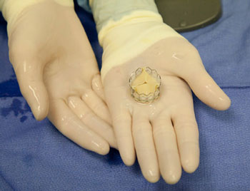
Percutaneous aortic valve replacement is being performed at the leading cardiac surgical centers around the world. This new technique does not require opening the chest, stopping the heart, or using the heart-lung machine to replace the aortic valve. The valve can be inserted through either an artery in the leg or a small incision between the ribs. This new valve is currently FDA approved for use in patients too old or sick for conventional surgery and likely to receive FDA approval in the near future for higher-risk patients with critical aortic stenosis.


As a pioneer in the field of aortic valve replacement using a catheter-based system that does not require use of the heart lung machine, stopping the heart, or opening the chest, Dr. Dewey has the distinction of performing the first transapical aortic valve replacement utilizing this breakthrough technology in the United States. He is recognized as one of the most experienced surgeons in the world with this technique. Internationally known as a leader in the field of valvular heart disease, he has trained surgeons in Europe, Canada, Australia, and the United States in both catheter-based aortic valve replacement as well as minimally invasive surgical techniques.
Dr. Todd Dewey is a member and active researcher at the Cardiopulmonary Research and Technology Institute (CRSTI) located at Medical City Dallas Hospital.
CRSTI is dedicated to turning tomorrow's medical science into today's treatments and cures, ensuring that the highest quality of care is available to the community. CRSTI has accomplished remarkable strides in its effort to develop and promote state-of-the-art therapies and techniques on behalf of those affected by heart and lung disease.
Lung Cancer Surgery Expert - A Q&A With Dr. Magee
Lung cancer causes more deaths than the next three most common cancers combined (colon, breast and prostate). It accounts for almost 30 percent of all cancer deaths, approximately 15 percent of all cancer diagnoses. In 1987, it surpassed breast cancer to become the leading cause of cancer deaths in women. Despite a continued decline in smoking, lung cancer is still increasing among non-smokers as well as women and can advance to a late stage without any symptoms.
Finding the right specialist is critical:
- A thoracic surgeon specializes in treating lung cancer and other diseases of the chest, other than the heart. This is a “super specialist” beyond general and cardiothoracic surgery.
- A thoracic surgeon has had years of additional training in their area of expertise (lung cancer). Therefore, you are in the hands of an expert in their field.
- Your lung cancer is more likely to be staged correctly - (Stages 1, 2, 3 or 4) if you consult with a thoracic surgeon, which in turn is critical in receiving the correct treatment based on stage.
- Your long-term prognosis is more likely to be positive if you are treated by a surgeon who specializes in lung cancer. That is a thoracic surgeon.
- An experienced thoracic surgeon will more likely use minimally-invasive techniques with greater results due to his or her specialized training and expertise for your lung cancer.
- Studies have shown that risks are lower and cure rates higher when lung cancer procedures are performed by thoracic surgeons compared to general or cardiothoracic surgeons.